
apr 21, 2023
Het scherpste beeld van een brein maak je met geluid
Hoe breng je de kleine bloed en haarvaten van een brein haarscherp in beeld? Een team van onderzoekers aan de TU/e in samenwerking met Philips Research Ultrasound, gingen de uitdaging aan om deze vraag te beantwoorden. Het team slaagde erin een rattenbrein zo gedetailleerd in kaart te brengen, dat ze de tweede plek wonnen in de ULTRA-SR Challenge (Ultrasound Localisation and TRacking Algorithms for Super Resolution). Hoe ze dat deden, presenteerden ze tijdens een e/MTIC webinar voor e/MTIC partners en PhD studenten.
Eindhoven MedTech Innovation Center
Het onderzoek werd uitgevoerd binnen het samenwerkingsverband e/MTIC, het Eindhoven MedTech Innovation Center. Hierin werken de TU/e, het Catharina Ziekenhuis, Maxima Medisch Centrum (MMC), het Centrum voor Slaapgeneeskunde Kempenhaeghe en Philips samen aan diverse onderzoeken rondom cardiovasculaire, perinatale en slaap geneeskunde. In totaal werken er 100 PhD-studenten binnen het samenwerkingsverband om de implementatie van nieuwe gezondheidstechnologie te versnellen. Deze PhD studenten en partnerbedrijven werden uitgenodigd om de e/MTIC webinar “What does a brain sound like?” bij te wonen.
Beeldvorming met behulp van microbelletjes
Met medische beeldvorming, of radiologie, kunnen we gezondheidsproblemen in het lichaam in beeld brengen. Om de kleinste bloedvaten in een super hoge resolutie in beeld te kunnen brengen, kan men gebruik maken van een opkomende techniek genaamd Ultrasound Localization Microscopy (ULM)- een vorm van echografie. Hierbij worden microbelletjes ter grootte van rode bloedcellen in de bloedbaan gebracht, en wordt door middel van de weerkaatsing van geluidsgolven vastgesteld hoe deze vaten eruitzien. Echter, in delen van het lichaam waar de weerkaatsing van geluidsgolven belemmerd wordt, is beeldvorming onduidelijk.
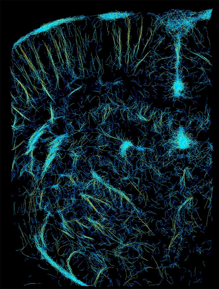
Een conventioneel ULM Super Resolution Ultrasound van een rattenbrein
De Ultrasound- Super- Resolution Challenge
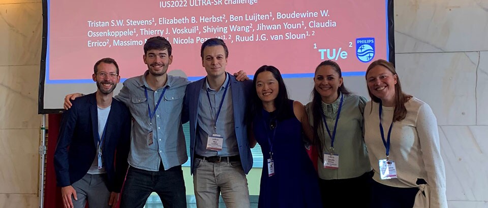
Het Institute of Electrical and Electronics Engineers (IEEE) nodigde daarom teams uit om het scherpste beeld te ontwikkelen in de Ultra Super Resolution Challenge. Het team van de TU/e en Philips werkte negen maanden aan de uitdaging om het scherpste beeld van een rattenbrein te vormen. Ze presenteerde hun model tijdens de Ultra SR- challenge in Venetië, waar 15 teams deelnamen in de categorie in-vivo beeldvorming van humane lymfeklieren en rattenbrein. “Onze doelstelling was om een robuust en betrouwbaar model te ontwikkelen die ook de onduidelijke delen van het brein in kaart kunnen brengen. Met behulp van data uit delen van het brein met een lage concentratie microbubbels, konden we ons model trainen in gebieden met een hoge concentratie bubbels; waar conventionele methodes vaak slecht presteren,” aldus Tristan Stevens, PhD student aan de TU/e Electrical Engineering. Het beeld leverde hen een tweede plek op in de challenge.
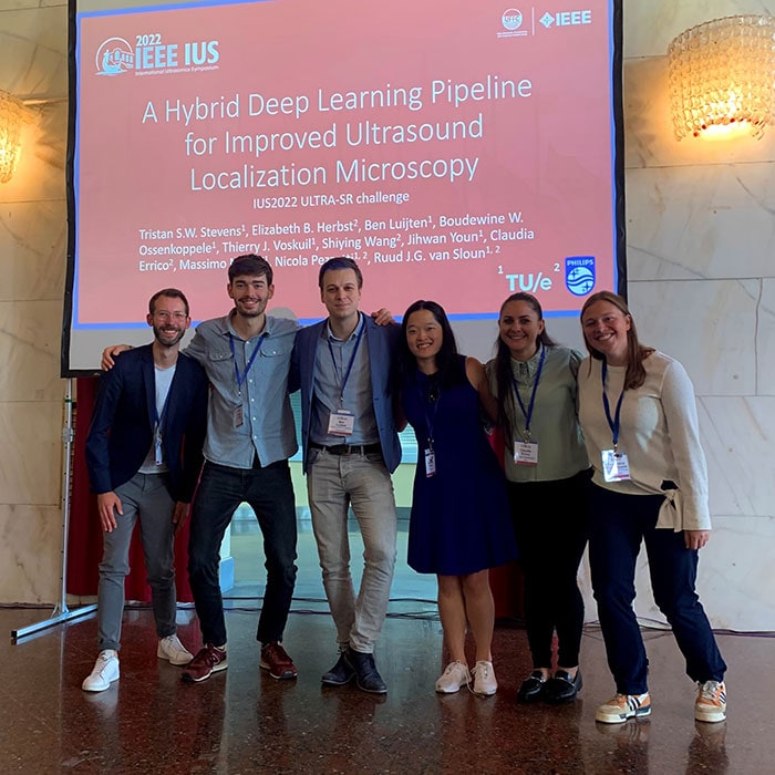
Het team (vlnr): Ruud van Sloun, Tristan Stevens, Ben Luijten, Elizabeth Herbst, Claudia Errico, Boudewine Ossenkoppele. Ontbrekend op de foto: Jihwan Youn, Thierry Voskuil, Shiying Wang, Nicola Pezzotti, Massimo Mischi
Beter beeld voor betere diagnoses
Met deze super resolutie beelden van de bloedvaten, kunnen artsen in de toekomst beter en eerder diagnoses stellen. “Naast een betere diagnose, kunnen de beelden ondersteunen in het eerder vaststellen van een letsel, zoals de voorstadia van leverkanker,” aldus Thanasis Loupas, Principal Clinical Scientist bij Philips. “Wanneer de micro vasculaire structuur verandert in de voorstadia van kanker, kunnen we dit mogelijk in kaart brengen met deze super resolutie beelden.”
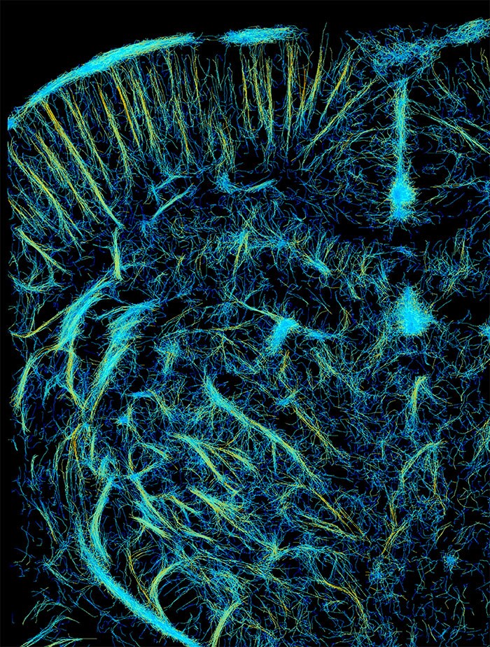
Het verbeterde Super-Resolution beeld van het e/MTIC team
E/MTIC samenwerking: de ware winst
“De samenwerking tussen universiteiten en het bedrijfsleven in dit veld is kritisch voor de ontwikkeling van ultrasound technologie,” aldus Sjoerd Mentink, Director Public Private Partnerships bij Philips. Thanasis vult aan: “Deze prijs is een bewijs dat wanneer verschillende perspectieven vanuit de academische wereld en het bedrijfsleven bij elkaar komen, er mooie dingen gebeuren. Voor mij is dat de echte winst van het team.”
The sharpest image of a brain is made with sound
How can you visualize small blood vessels and capillaries of a brain in a super-sharp image? A team from the Technical University Eindhoven (TU/e) together with Philips Research Ultrasound, managed to map a rat brain in such detail that they won second place in the ULTRA-SR Challenge (Ultrasound Localization and TRacking Algorithms for Super Resolution). They explained how they did this, during an e/MTIC webinar for e/MTIC partners and PhD students.
Eindhoven MedTech Innovation Center
The research was conducted within the partnership e/MTIC, the Eindhoven MedTech Innovation Center. In this partnership, the TU/e, Catharina Hospital, Maxima Medisch Centrum (MMC), Kempenhaeghe Center for Sleep Medicine and Philips work together on various research projects around cardiovascular, perinatal and sleep medicine. A total of 100 PhD students are working within the partnership to accelerate the implementation of new health technology. The e/MTIC PhD students and partner companies were invited to attend the e/MTIC webinar "What does a brain sound like?".
Imaging using microbubbles
Medical imaging, or radiology, allows us to visualize health problems in the body. To create a high-resolution image of our smallest blood vessels, the emerging technique called Ultrasound Localization Microscopy (ULM)-a form of ultrasound- can be used. This involves bringing microbubbles the size of red blood cells into the bloodstream, and using the reflection of sound waves to determine what these vessels look like. However, in parts of the body where the reflection of sound waves is obstructed, imaging is unclear.
A conventional super resolution ultrasound image of a rat brain
The Ultrasound- Super- Resolution Challenge
The Institute of Electrical and Electronics Engineers (IEEE) invited teams to develop the sharpest image in the Ultra Super Resolution Challenge. The TU/e and Philips team worked for nine months on the challenge to form the sharpest image of a rat brain. They presented their model at the Ultra SR challenge in Venice, where 15 teams participated in the category of in-vivo imaging of human lymph nodes and rat brain. "Our objective was to develop a robust and reliable model that can also map the unclear parts of the brain. Using data from parts of the brain with a low concentration of microbubbles, we were able to train our model in areas with a high concentration of bubbles; where conventional methods often perform poorly," said Tristan Stevens, PhD student at TU/e Electrical Engineering.
The team (fltr): Ruud van Sloun, Tristan Stevens, Ben Luijten, Elizabeth Herbst, Claudia Errico, Boudewine Ossenkoppele. Missing in the picture: Jihwan Youn, Thierry Voskuil, Shiying Wang, Nicola Pezzotti, Massimo Mischi
Better image for better diagnoses
The image earned them second place in the challenge. With these super resolution images of blood vessels, doctors will be able to make better and earlier diagnoses in the future. "In addition to better diagnosis, the images may support earlier identification of a lesion, such as the pre-stages of liver cancer," said Thanasis Loupas, Principal Clinical Scientist at Philips. "When micro vascular structure changes in the pre-stages of cancer, we may be able to map this with these super resolution images."
The improved Super-Resolution image of the e/MTIC team
e/MTIC collaboration: the true benefit
"University-industry collaboration in this field is critical for the development of ultrasound technology," said Sjoerd Mentink, Director of Public Private Partnerships at Philips. Thanasis adds: "This award is proof that when different perspectives from academia and industry come together, great things happen. For me, that is the true benefit of the team."
Deel op social media
Onderwerpen
Contact

Tommie Dijstelbloem
Woordvoerder Philips Benelux Tel: +31 6 19 28 83 20
You are about to visit a Philips global content page
Continue(Meer) gerelateerd nieuws
-
![Philips Girl’s Day 2024: ‘Geef jezelf gelijke kansen’]()
april 16, 2024


