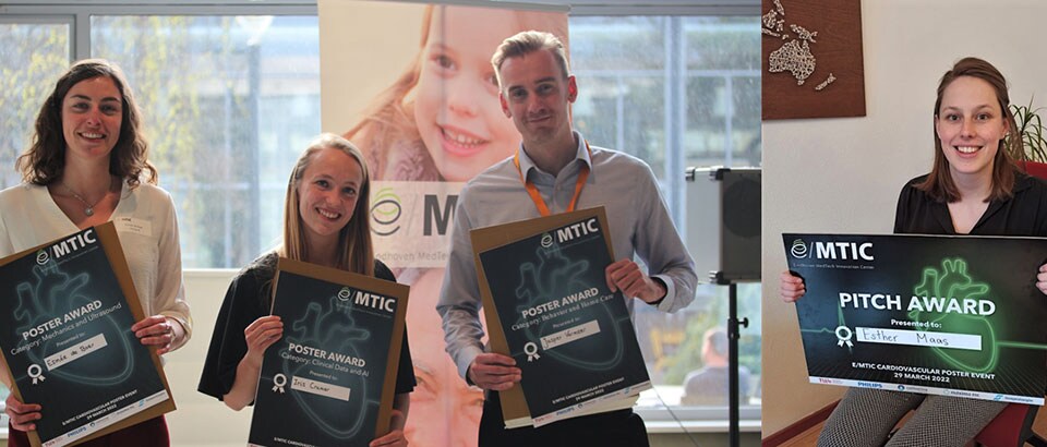
Patiënten met een verwijding in de buikslagader lopen een risico dat het bloedvat gaat scheuren; waarbij patiënten in sommige gevallen kunnen overlijden. Esther Maas, PhD-studente bij de Technische Universiteit Eindhoven (TU/e) en het Catharina Ziekenhuis, werkt aan een innovatieve manier om het risico op een scheur beter in te schatten – en dus de behandeling effectiever te maken. Het onderzoek, en de manier waarop ze het wist te presenteren, leverde haar een prijs op tijdens het e/MTIC evenement Cardiovascular Medicine.
De implementatie van nieuwe gezondheidstechnologie versnellen
Esther’s onderzoek wordt uitgevoerd binnen het samenwerkingsverband e/MTIC, het Eindhoven MedTech Innovation Center. Hierin werken de TU/e, het Catharina Ziekenhuis, Maxima Medisch Centrum (MMC), het Centrum voor Slaapgeneeskunde Kempenhaeghe en Philips samen aan diverse onderzoeken rondom cardiovasculaire, perinatale en slaap geneeskunde. In totaal werken er honderd PhD-studenten binnen het samenwerkingsverband om de implementatie van nieuwe gezondheidstechnologie te versnellen.
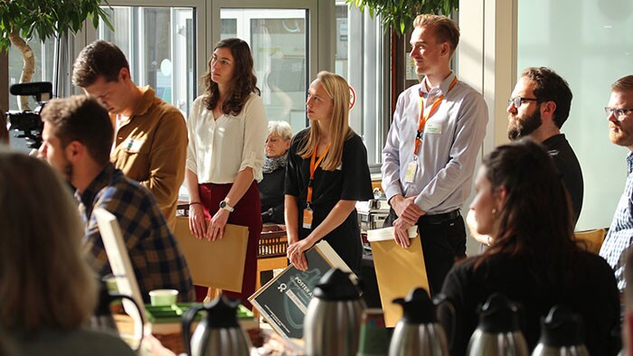
In het e/MTIC Poster Event - CardioVascular Medicine werden deelnemers uitgenodigd om ideeën uit te wisselen en met elkaar in contact te komen. “Voor mij een ideale gelegenheid om mijn onderzoek te toetsen bij mede-onderzoekers en de samenwerking op te zoeken met andere deelnemers.”

Maat bepalend in risico-inschatting
Bij een aneurysma is een deel van een bloedvat minimaal anderhalf keer zo wijd als normaal. Het komt het meest voor in de buikslagader, een abdominaal aorta aneurysma (AAA). Een AAA geeft meestal geen klachten en wordt daarom vaak bij toeval geconstateerd, maar als het te groot is of snel groeit, bestaat er een risico op een scheur in de bloedvatwand. Met mogelijk overlijden tot gevolg. Het is daarom belangrijk om de patiënt na de constatering van de verwijding te blijven monitoren. Wordt de verwijding te groot, dan komt de patiënt in aanmerking voor een operatie.
De grootte van een aneurysma is niet voor iedere patiënt een goede indicatie voor de noodzaak voor een operatie.
Esther Maas
PhD-student
Meer dan maat alleen
De grootte van het aneurysma is echter niet voor iedere patiënt een goede indicatie voor het risico op een scheur. “Zo kan een scheur ook optreden bij een kleiner aneurysma of kan een patiënt geen last ondervinden van een grotere variant,” aldus Esther. In haar onderzoek worden andere factoren onderzocht om het risico op een scheur te voorspellen.
“In ziekenhuizen wordt nu een 2D-echografie of een CT-scan gebruikt om de diameter van de buikslagader vast te stellen”, vervolgt Esther. “Met 4D Ultrasound zijn we nu in het Catharina Ziekenhuis in staat om naast de diameter, ook de vorm en elasticiteit van het aneurysma te meten. Vervolgens kunnen we op basis van deze factoren in een computermodel de krachten simuleren die in het lichaam op de vaatwand werken. Hoe groter de spanningen die hierdoor in de vaatwand ontstaan, hoe groter de kans op een scheur. Zo kunnen we uiteindelijk het risico voor de patiënt inschatten en in de toekomst de noodzaak tot opereren hierop afstemmen.”
In haar onderzoek maakt Esther gebruik van een nieuwe variant van een gangbare technologie. “Met behulp van 4D ultrasound is het mogelijk de gehele vorm en de beweging van het aneurysma te bekijken. Daarnaast is het in de klinische toepassing van belang dat we een toegankelijke en kosten-efficiënte technologie gebruiken”, legt Esther uit.
Hoe knapt een ballon?
Om haar onderzoek treffend uit te leggen aan haar publiek, gebruikt Esther de analogie van het knappen van een ballon en de factoren die daarbij van belang zijn. Voor de juryleden van het e/MTIC cardiovascular medicine pitch was dit één van de redenen om Esther de titel ‘Beste Pitch’ toe te kennen.
“Esther wist met deze treffende introductie op een eenvoudige en herkenbare manier haar onderzoek uit te leggen aan het publiek”, aldus Sjoerd Mentink, Program Manager voor e/MTIC bij Philips en jurylid. “Daarnaast maakt Esther in haar onderzoek gebruik van een bekende meetmethode”, vult dr. Fokke van Meulen, Postdoc bij de TU/e en Kempenhaeghe hem aan. “Dat betekent dat de resultaten van haar onderzoek in potentie snel een belangrijke en impactvolle verandering in de dagelijkse klinische praktijk teweeg kunnen brengen.” Naast Sjoerd Mentink en Fokke van Meulen, bestond de jury uit Myrthe van der Ven (TU/e en Máxima Medisch Centrum) en Henning Maass (Philips).
Esther maakt in haar onderzoek gebruik van een bekende meetmethode. Dat betekent dat de resultaten van haar onderzoek in potentie snel een belangrijke en impactvolle verandering in de dagelijkse klinische praktijk teweeg kunnen brengen.
dr. Fokke van Meulen
Postdoc bij de TU/e en Kempenhaeghe

“By using computer models, we can better predict the risk of a rupturing blood vessel”
Patients with a dilated abdominal artery are at risk of a blood vessel rupture; in extreme cases patients may die. Esther Maas, PhD student at Eindhoven University of Technology (TU/e) and Catharina Hospital, is working on an innovative way to better assess the risk of a rupture - and thus make the treatment more effective. The research, and the way she managed to present it, earned her an award at the e/MTIC event Cardiovascular Medicine.
Accelerating the implementation of new health technology
Esther's research is conducted within the partnership e/MTIC, the Eindhoven MedTech Innovation Center. In this partnership, TU/e, Catharina Hospital, Maxima Medical Center (MMC), the Center for Sleep Medicine Kempenhaeghe and Philips are working together on various studies related to cardiovascular, perinatal and sleep medicine. A total of 100 PhD students are working within the partnership to accelerate the implementation of new health technology.
In the e/MTIC Poster Event - CardioVascular Medicine, participants were invited to exchange ideas and connect with each other. "For me, an ideal opportunity to test my research with fellow researchers and seek collaboration with other participants."

Size determines risk assessment
In an aneurysm, part of a blood vessel is at least one and a half times wider than normal. It is most common in the abdominal artery, an abdominal aortic aneurysm (AAA). An AAA usually does not cause any symptoms and is therefore often detected by accident, but if it is too large or grows rapidly, there is a risk of a tear in the blood vessel wall. With possible death as a result. It is therefore important to continue to monitor the patient after the dilatation is detected. If the dilatation becomes too large, the patient will be considered for surgery.
The size of an aneurysm might not be a good indication of the need for surgery for every patient.
Esther Maas
PhD student
More than size
However, the size of the aneurysm is not a good indication of the risk of a rupture for every patient. "For example, a tear may also occur with a smaller aneurysm, or a patient may not suffer from a larger variant," says Esther. Her research is examining other factors to predict the risk of a tear.
"In hospitals, a 2D ultrasound or CT scan is now used to determine the diameter of the abdominal artery," Esther continued. "With 4D Ultrasound, we are now able at Catharina Hospital to measure not only the diameter, but also the shape and elasticity of the aneurysm. Based on these factors, we can then simulate the forces that act on the vessel wall in the body in a computer model. The greater the stress this creates in the vessel wall, the greater the likelihood of a rupture. This ultimately allows us to assess the risk to the patient and in the future to adjust the need for surgery accordingly."
In her research, Esther uses a new variation of a common technology. "Using 4D ultrasound, it is possible to view the entire shape and movement of the aneurysm. In addition, it is important in the clinical application that we use an accessible and cost-effective technology," explains Esther.
How to snap a balloon?
To aptly explain her research to her audience, Esther uses the analogy of a balloon snapping and the factors involved. For the judges of the e/MTIC cardiovascular medicine pitch, this was one of the reasons for awarding Esther the title of "Best Pitch”.
"Esther managed to explain her research to the audience in a simple and recognizable way with this striking introduction," said Sjoerd Mentink, Program Manager for e/MTIC at Philips and member of the jury. "In addition, Esther uses a well-known measurement method in her research," adds Dr. Fokke van Meulen, Postdoc at TU/e and Kempenhaeghe. "This means that the results of her research have the potential to quickly bring about an important and impactful change in daily clinical practice." In addition to Sjoerd Mentink and Fokke van Meulen, the jury included Myrthe van der Ven (TU/e and Máxima Medisch Centrum) and Henning Maass (Philips).
Esther uses a known measurement method in her research. This means that the results of her research have the potential to quickly bring about an important and impactful change in daily clinical practice.
dr. Fokke van Meulen
Postdoc at TU/e and Kempenhaeghe


